Many pregnant women thinking about getting an early ultrasound have questions. We want to answer your questions so you know what to expect.
A few things to know about early ultrasound scans:
- Ultrasounds at Pacific Pregnancy Clinic are free
- Ultrasounds are safe
- We can help you prepare – and you do not have to go alone
- How early ultrasounds work and what they look like
- Common reasons for an early ultrasound
If you have more questions after reading this article, we would love to talk with you. Schedule an appointment with us, below.

Are You Pregnant?
Find out for sure with a free test at Pacific Pregnancy Clinic
Get My Free Test
ULTRASOUNDS AT Pacific Pregnancy Clinic ARE FREE, EVEN IF YOU DO NOT HAVE INSURANCE.
Thanks to our generous donors, we are able to provide you with a free ultrasound. You do not need insurance. We provide this as a service to you and your unborn baby. Our pregnancy center is a nonprofit organization, and we are here to support you. We keep your information completely confidential and do not profit from any decision you make. For more information about pregnancy centers, click here. (1)
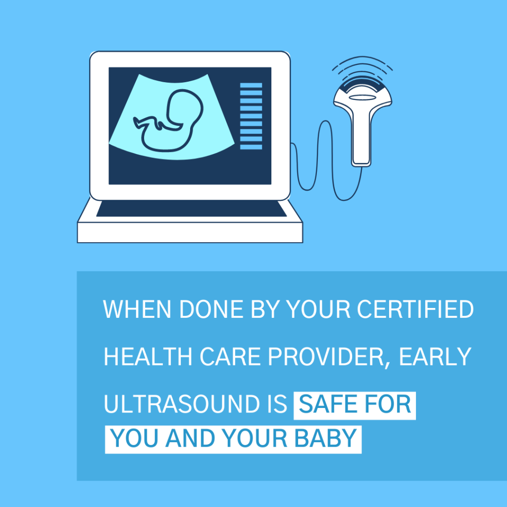
WHEN DONE BY A CERTIFIED HEALTH CARE PROVIDER, ULTRASOUNDS ARE SAFE FOR YOU AND YOUR BABY.
Early ultrasounds are medical diagnostic procedures. An ultrasound machine is safe when used by a certified health care provider. If you schedule a prenatal ultrasound with us, you and your baby will be free of any dangerous risks. (2)
We understand if you are anxious to see your ultrasound image and we are happy to show you images from your ultrasound scan. Keeping that in mind, the reason for an early ultrasound is to help you know about your health and your developing baby. For that reason, we encourage you to view this procedure as a medical process that serves your health and your baby’s health. There is more information about common reasons for early ultrasounds below.


PREPARING FOR YOUR ULTRASOUND.
There are a few things you might find helpful to know before scheduling an appointment with us. The first thing to know is that we may ask you to drink fluids and avoid urinating for a specific amount of time before the procedure. (More about this in the next section.)
We also want you to know that you do not have to come to your appointment alone. If you would like, you may bring your husband or boyfriend, a parent, or another supporter. It may be helpful for them to be aware of how things are developing with your pregnancy as well.
HOW EARLY ULTRASOUNDS WORK AND WHAT THEY LOOK LIKE.
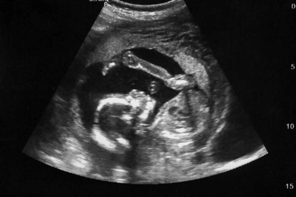
A fetal ultrasound, also called a sonogram, produces images of your baby in the womb by using high-frequency sound waves.
Early ultrasounds are done in the first 14 weeks of pregnancy and show images of the baby’s early development stages. (3) Routine ultrasound images are typically black and white. They are usually somewhat fuzzy but are detailed enough to show us what we need to know about fetal growth. For this reason, it may not be easy for you to identify exactly what you are seeing on the screen. We’ll help you understand what we’re seeing and what it means. We will also give you the option of keeping a few printed images from the scan.
There are a few things you will notice when you look at the ultrasound screen. You will see a white image of your baby and umbilical cord, against a dark background. Your doctor will learn a lot of important information about your developing baby by observing these images.
For example, a 7-week old baby is just the size of a blueberry, but already has developed limb buds, outer ears, and nearly complete eyelids. At this stage, your baby also has an increased heart rate since the last week and cells that are developing muscles and a spinal column. (5)
We share more details about what we can learn together after we cover the two types of ultrasounds.
THERE ARE TWO TYPES OF EARLY ULTRASOUNDS: ABDOMINAL AND VAGINAL.
The transabdominal or standard ultrasound is what you’re probably picturing. (4) To prepare for transabdominal ultrasounds, we ask you to drink a few glasses of water a couple of hours before the procedure. This is because a full bladder helps the high frequency sound waves move more easily, to help provide a clear picture. (6)
This is a painless procedure and it usually takes about 20 minutes. To begin, we will ask you to lay on your back on an examining table. Next, we will apply a gel to your abdomen and move a scanner over it. The scanner is a small, hand-held device that is connected to a screen which instantly shows images.
Vaginal Ultrasounds
Another type of ultrasound is transvaginal ultrasound. These are usually done for the early stages of pregnancy, or when the images from a transabdominal ultrasound are not quite clear enough. We do not provide this type of ultrasound, but it may be helpful to know about it because it is common.
For this procedure, your health care provider will likely ask you to change into a gown and undress from the waist down. Next, you will lie down on an examining table and place your feet in stirrups.
For this process, your health care provider will use a small, slender scanner. The scanner is shaped like a wand. It is covered with a plastic sheath and lubricated before being placed into your vagina. The process is also about 20 minutes long and may cause some discomfort, but shouldn’t be painful. (3)
Both types of early ultrasounds are for early pregnancy, in your first trimester. To learn more about additional ultrasounds in the second trimester or third trimester of pregnancy, click here. (7)
COMMON REASONS FOR AN EARLY ULTRASOUND.
One of the most common reasons for an early ultrasound is to confirm pregnancy, which your doctor can also confirm with a blood test. Other reasons include: checking if there is more than one baby and determining your baby’s gestational age, health, and location. Gestational age estimates the weeks of gestation to determine your baby’s due date.
When it comes to your baby’s health, we will be checking on your baby’s heartbeat, muscle tone, movement, and determine if there are any birth defects. (3)
Location is something we will look for, because in certain cases a baby may be developing outside the main cavity of the uterus. This is called an ectopic pregnancy. Most ectopic pregnancies are in the fallopian tube and you can learn more here. (3) Additionally, we will be able to examine the health of your ovaries and uterus. (2)

LIMITS OF EARLY ULTRASOUND
Your ultrasound test will be 2D, not 3D images or 4D ultrasound. However, there is a lot of useful information we can learn together from your pregnancy ultrasound.
It will also be helpful for you to know about a few things that an early ultrasound cannot determine. First, it cannot determine your baby’s sex. This is because early ultrasounds are done in the first trimester, and a baby’s gender can only be determined after the second trimester, after 18 to 21 weeks of pregnancy. (3)
Second, ultrasounds in the first six to eight weeks of pregnancy cannot determine the presence of Down syndrome. Doctors don’t perform a nuchal translucency ultrasound for Down syndrome until 14–20 weeks gestation, and then only if a past screening test showed a problem. (3) For other types of ultrasounds and how they work, click here. (2)
What we learn about your health, and the health of your baby will help you make decisions that are best for you both. This is the first step in the process and we would love to go on the journey with you.
